Home

Christentum Vier Richtung gradient echo mri brain Timer Genius Geldüberweisung

EPOS™
EPOS™

Cognitive dysfunction in patients with cerebral microbleeds on T2*-weighted  gradient-echo MRI
Cognitive dysfunction in patients with cerebral microbleeds on T2*-weighted gradient-echo MRI

Gradient echo T1 (left: TR, 311 ms; TE, 2.5 ms; section thickness, 5... |  Download Scientific Diagram
Gradient echo T1 (left: TR, 311 ms; TE, 2.5 ms; section thickness, 5... | Download Scientific Diagram

11 Top Brain MRI ideas | mri, protected health information, brain

Gradient Echo MRI
Gradient Echo MRI

Brain MRI. Flair sequence (a) and gradient echo sequence(b). Initial... |  Download Scientific Diagram
Brain MRI. Flair sequence (a) and gradient echo sequence(b). Initial... | Download Scientific Diagram
Automated detection of cerebral microbleeds on T2*-weighted MRI |  Scientific Reports
Automated detection of cerebral microbleeds on T2*-weighted MRI | Scientific Reports

High diagnostic value of gradient echo sequence in isolated cortical vein  thrombosis
High diagnostic value of gradient echo sequence in isolated cortical vein thrombosis

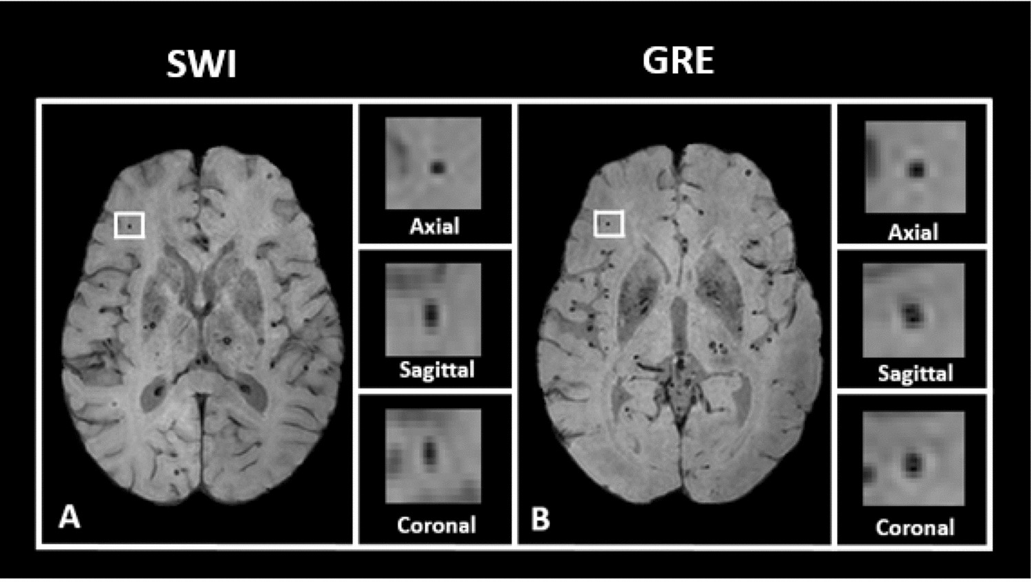
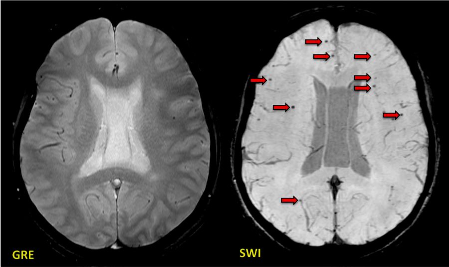
Superiority of SWI over GRE - Questions and Answers ​in MRI

GRE v SE - Questions and Answers ​in MRI

Susceptibility weighted imaging - Wikipedia

MRI Protocols: Gradient echo sequence use in MRI BRAIN

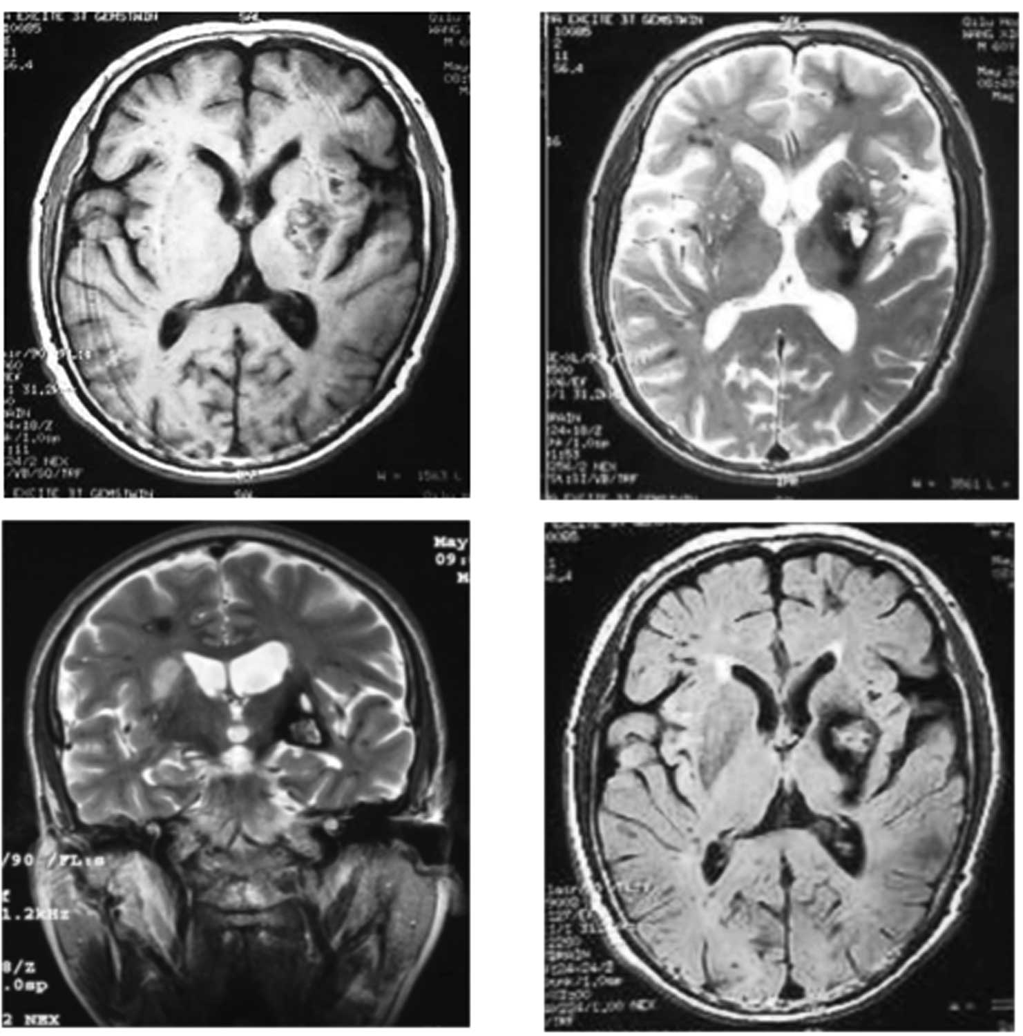
MRI BRAIN with T1 T2 (top left and right) and gradient echo (GRE,... |  Download Scientific Diagram
MRI BRAIN with T1 T2 (top left and right) and gradient echo (GRE,... | Download Scientific Diagram

The value of T2*-weighted gradient echo imaging for detection of familial  cerebral cavernous malformation: A study of two families
The value of T2*-weighted gradient echo imaging for detection of familial cerebral cavernous malformation: A study of two families

MRI detection of hypointense brain lesions in patients with multiple  sclerosis: T1 spin-echo vs. gradient-echo. | Semantic Scholar
MRI detection of hypointense brain lesions in patients with multiple sclerosis: T1 spin-echo vs. gradient-echo. | Semantic Scholar

Hypointensities in the Brain on T2*-Weighted Gradient-Echo Magnetic  Resonance Imaging - ScienceDirect
Hypointensities in the Brain on T2*-Weighted Gradient-Echo Magnetic Resonance Imaging - ScienceDirect

Diagnostic value of T2*-weighted gradient-echo MRI for segmental evaluation  in cerebral venous sinus thrombosis. | Semantic Scholar
Diagnostic value of T2*-weighted gradient-echo MRI for segmental evaluation in cerebral venous sinus thrombosis. | Semantic Scholar
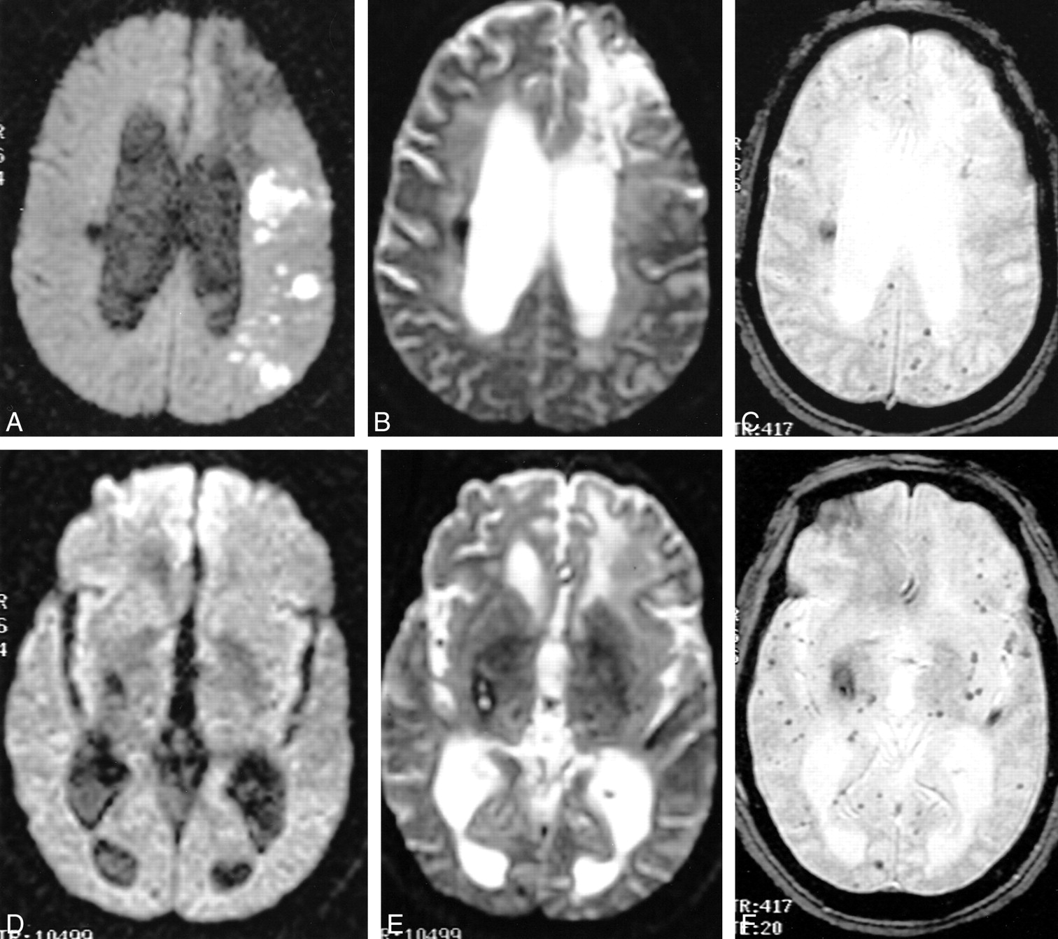
Detection of Intracranial Hemorrhage: Comparison between Gradient-echo  Images and b0 Images Obtained from Diffusion-weighted Echo-planar Sequences  | American Journal of Neuroradiology
Detection of Intracranial Hemorrhage: Comparison between Gradient-echo Images and b0 Images Obtained from Diffusion-weighted Echo-planar Sequences | American Journal of Neuroradiology

Gradient echo imaging - Markl - 2012 - Journal of Magnetic Resonance Imaging  - Wiley Online Library
Gradient echo imaging - Markl - 2012 - Journal of Magnetic Resonance Imaging - Wiley Online Library

Spoiled GRE - Questions and Answers ​in MRI

T1, T2, FLAIR, and Gradient Echo pulse sequences. - YouTube

Isolated foot drop due to subcortical hemorrhage | SpringerLink

Detection of Intracranial Hemorrhage: Comparison between Gradient-echo  Images and b0 Images Obtained from Diffusion-weighted Echo-planar Sequences  | American Journal of Neuroradiology
Detection of Intracranial Hemorrhage: Comparison between Gradient-echo Images and b0 Images Obtained from Diffusion-weighted Echo-planar Sequences | American Journal of Neuroradiology

Improved T2* Imaging without Increase in Scan Time: SWI Processing of 2D Gradient  Echo | American Journal of Neuroradiology
Improved T2* Imaging without Increase in Scan Time: SWI Processing of 2D Gradient Echo | American Journal of Neuroradiology

MRI brain (Gradient echo images) showed acute hemorrhages... | Download  Scientific Diagram
MRI brain (Gradient echo images) showed acute hemorrhages... | Download Scientific Diagram

MRI of brain T2-weighted axial gradient echo sequence. a At diagnosis.... |  Download Scientific Diagram
MRI of brain T2-weighted axial gradient echo sequence. a At diagnosis.... | Download Scientific Diagram